
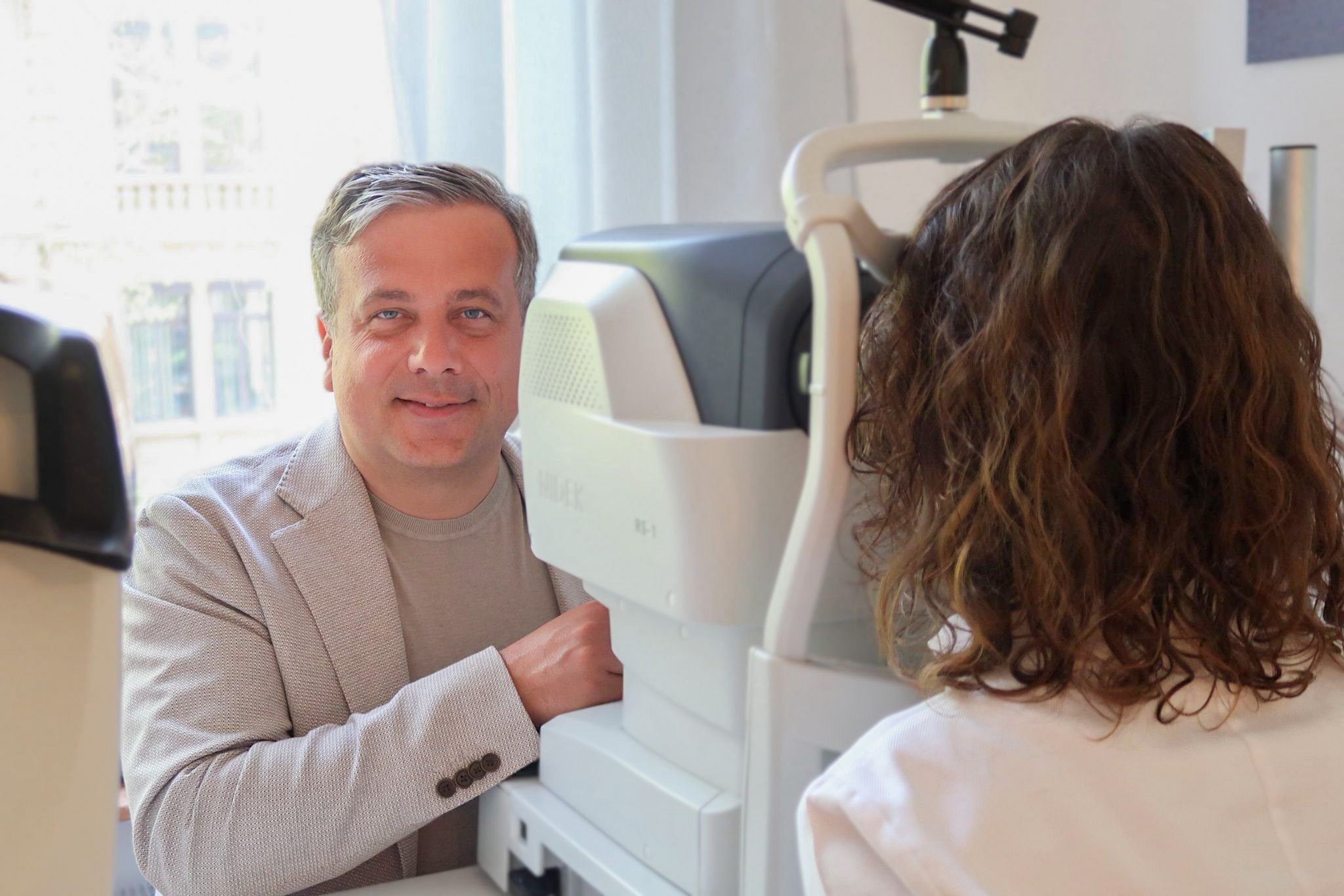
The Glaucoma is an insidious, often painless disease which, if left untreated, can lead to irreversible blindness can lead. The Glaucoma screening In our Frankfurt practice, one of the most important preventative measures to protect your eyesight is therefore... Dr. Mareen Schmidt & Dr. Sebastian Schmidt in the Westend district focus on... State-of-the-art, gentle diagnostics, this insidious disease to recognize at an early stage and secure your eyesight in the long term.
Glaucoma is not a single disease, but a group of eye diseases that have one thing in common: They lead to progressive and irreversible damage to the optic nerve. The optic nerve is the „data cable“ that transmits all visual impressions from the eye to the brain. If the millions of nerve fibers that make it up are damaged, gaps occur in the visual field. As these losses typically begin at the edge of the visual field and central vision remains intact for a long time, those affected often only notice the disease at a very late stage. An effective Glaucoma screening is therefore the only way to stop this „silent thief of sight“ in time.
Comprehensive glaucoma screening in our Frankfurt practice goes far beyond a simple measurement. It consists of several complementary components, which together provide a precise picture of the health of your optic nerve and your individual risk.
Intraocular pressure measurement (tonometry): Elevated intraocular pressure is the most important risk factor for the development of glaucoma. We carry out this measurement gently and contact-free, without having to touch your eye directly. Our devices are FDA-approved.
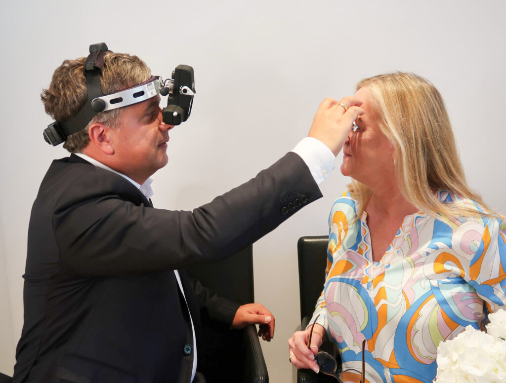
Assessment of the optic nerve head (ophthalmoscopy): Dr. Mareen Schmidt uses a special microscope to examine the optic nerve head, the point at which the optic nerve exits the eye. Typical glaucoma changes, such as an increasing hollowing („excavation“), can be detected here at an early stage.
Corneal thickness measurement (pachymetry): The thickness of your cornea has an influence on the accuracy of the intraocular pressure measurement. By measuring the corneal thickness, we can individually correct the measured pressure value and assess it even more precisely.
Papillary OCT and OCT-A (optical coherence tomography and angiography): These high-resolution, non-invasive imaging techniques allow precise measurement of the nerve fiber layer at the optic nerve head (optic disc) and in the area of the macula. The Papilla OCT detects even the finest, early nerve fiber loss before it becomes visible in the visual field or during ophthalmoscopy. The OCT-A provides additional information about the blood supply to the optic nerve.
Visual field examination (perimetry): This functional examination measures whether there are already losses in your peripheral or central vision. As glaucoma damage often occurs late in the visual field, this examination is used to document and monitor the progression of advanced disease.
Interdisciplinary approach with internists for normal pressure glaucoma: In patients with normal pressure glaucoma, where the intraocular pressure is not elevated, vascular or systemic causes (e.g. blood pressure fluctuations, circulatory disorders) may play a role. In these cases, we work closely with experienced internists to identify and treat risk factors outside the eye.
Although a Glaucoma screening is generally recommended for every adult over the age of 40, there are certain risk groups for whom earlier and more regular check-ups are particularly important.
The optic nerve is more susceptible to pressure damage in very short-sighted eyes.
The risk increases significantly from the age of 40.
The main risk factor.
If your parents or siblings have glaucoma, your personal risk is significantly increased.
The following table gives you an overview of the most important procedures and their respective contribution to the diagnosis.
Procedure | What is measured/assessed? | Why is it important for glaucoma screening? |
Tonometry | Intraocular pressure. | Identifies the most important treatable risk factor. |
Ophthalmoscopy | The appearance and shape of the optic disc. | Enables direct visual detection of typical glaucoma-related damage. |
Pachymetry | The thickness of the cornea. | Improves the accuracy of intraocular pressure measurements. |
OCT of the optic nerve | The exact thickness of the nerve fiber layer. | It detects the earliest signs of structural damage—often years before other tests—and enables precise monitoring of the condition's progression. |
Glaucoma screening
The Glaucoma screening is based on a combination of various specific tests, each of which provides different but important information for the diagnosis and monitoring of the disease.
Measurement and Evaluation
The Tonometry is used to measure the Intraocular pressure – the most important treatable risk factor for glaucoma. Since elevated intraocular pressure can lead to damage to the optic nerve, this measurement is essential.
At the same time, the Ophthalmoscopy the ophthalmologist the direct visual assessment of the The appearance and shape of the optic disc. This study is crucial because it Detection of typical, pre-existing glaucoma damage on the nerve.
The ophthalmology professional associations recommend regular Glaucoma screening For everyone aged 40 and older. If risk factors are present—particularly a family history of the condition—the first screening should take place earlier. Starting screenings early is the best protection.
No, none of the studies conducted as part of the Glaucoma screening is painful. Modern intraocular pressure measurement is performed using a gentle puff of air and is non-contact. The OCT examination is also a purely non-invasive scanning procedure in which you simply look at a fixation light. Our gentle approach is part of our commitment to quality.
The recommended interval for the Glaucoma screening It depends on your age and your individual risk profile. If your test results are normal and your risk is low, a two-year interval may be sufficient. If you have increased risk factors or borderline test results, annual checkups are recommended to detect any changes early.
Yes, that's a very important point. There is a form of glaucoma known as "normal-tension glaucoma" in which the optic nerve is damaged even though intraocular pressure is within the normal statistical range. For this reason, measuring pressure alone is not sufficient for a reliable Glaucoma screening is not sufficient. The evaluation of the optic nerve is always the decisive factor.
A notable finding in the Glaucoma screening This is no reason to panic, but rather an opportunity to take action in a timely manner. Dr. Mareen Schmidt will discuss the findings with you in detail. Typically, a visual field test (perimetry) is then performed to check for any existing functional deficits. If the diagnosis is confirmed, treatment with eye drops can, in the vast majority of cases, stop or significantly slow the progression of the disease.
The most important step is to regularly participate in the Glaucoma screening, since you cannot feel the condition yourself. A healthy lifestyle that includes regular exercise and good management of blood pressure and blood sugar can support blood flow to the optic nerve. However, early detection by an ophthalmologist remains the key factor in protecting your vision.
For a comprehensive evaluation of the optic disc as part of the Glaucoma screening In many cases, dilating the pupil with medication is beneficial. Whether this is necessary for you depends on your individual anatomy. However, thanks to our state-of-the-art OCT technology, we can often perform a very precise analysis of the nerve fiber layer even without dilating eye drops.
These two diseases are often confused. Glaucoma, which occurs with the Glaucoma screening is a disease of the optic nerve. A cataract is a clouding of the lens of the eye that leads to blurred vision but can be completely cured by surgery. The damage caused by glaucoma, on the other hand, is irreversible.
In the practice of Dr. Mareen Schmidt & Dr. Sebastian Schmidt, you benefit from a Glaucoma screening at the highest level. We combine state-of-the-art technology such as optic nerve OCT with the many years of experience of a specialist. Our careful and gentle approach using the 5-S method ensures an examination that is as precise as it is comfortable. Your eyesight is in the best hands with us.
Generally not, you can drive afterwards, go to the office or do sports.




Check-ups, laser medicine, interdisciplinary ophthalmology

Retina, macular therapy, laser medicine, prevention & more
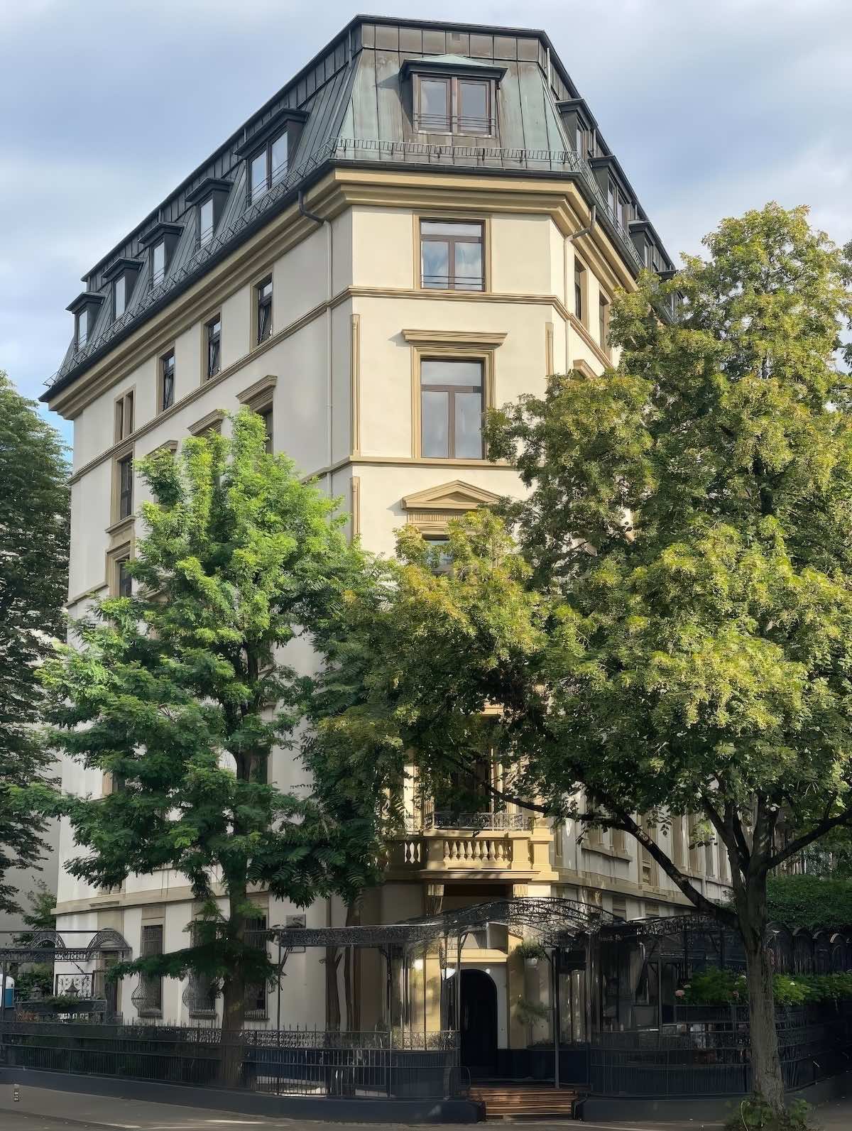
Address for the GPS:
Grüneburgweg 95 (corner of Liebigstraße), 60323 Frankfurt am Main.
Parking recommendations:
We recommend the following parking garages in the immediate vicinity:
Palmengarten parking garage: (Entrance via Siesmayerstraße). From there, it's about a 5-7 minute walk to the practice.
Campus Westend parking garage: (Entrance via Theodor-W.-Adorno-Anlage). Perfectly located for reaching the practice on foot in just a few minutes.
By bus (line 75):
Use the Bus route 75 and get off at the stop Eppsteiner Straße From there, it's only about a 2-3 minute walk (approx. 200 meters) along Grüneburgweg to the practice at the corner of Liebigstraße. Alternatively, you can use the bus stop. University Campus Westend to use.
By subway (U6 / U7):
Drive to the station Westend. Take the exit towards Feuerbachstraße/Liebigstraße. If you follow Liebigstraße straight ahead to the north, you will walk directly towards Villa Westend on Grüneburgweg (approx. 7-minute walk).
Alternative via the Palm Garden:
From the subway station Westend or the bus stop Palmengartenstrasse You can reach us after a short, pleasant walk past the Palm Garden.