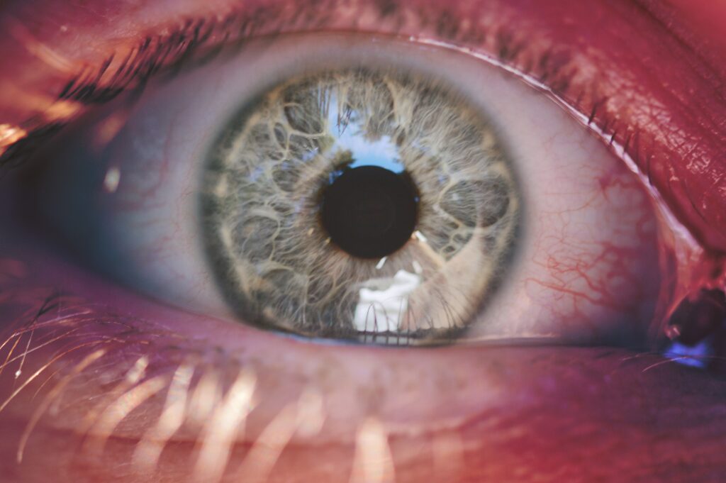
The retina (Retina) is the photosensitive layer in the back of the eye and functions like the Film in a camera. It consists of millions of photoreceptors (rods and cones), which absorb the incoming light stimuli and convert them into electrical signals. These signals are transmitted via the optic nerve to the brain, where they are processed into an image. A healthy retina is essential for the eyesight.
The Macula (also known as the „yellow spot“) is a tiny area in the center of the retina. It is the Most important region for our vision, as this is where the cone cells, which are responsible for Color vision and the Detailed, sharp vision are the most densely packed.
Central importance: Everything you focus on during direct vision - faces, letters, fine details - is captured by the macula.
Risk area: As the macula is constantly exposed to high levels of light and metabolic stress, it is particularly susceptible to age-related or disease-related changes.
As retinal and macular diseases often begin without symptoms, regular ophthalmologic check-ups are essential, especially if you have risk factors such as Age, diabetes, high blood pressure or high myopia show. In our practice, we use state-of-the-art diagnostics for early detection:
Your range of diagnostics
Retinal scanning (without wide-drop): Creation of Wide-angle digital photos of the retina for rapid screening, documentation and detection of peripheral changes.
Optical coherence tomography (OCT): High-resolution Sectional images of the macula and optic nerve for millimeter-precise visualization of swellings, fluid accumulations or nerve fiber loss.
OCT angiography (OCT-A): An innovative method for representation of the blood flow of the retina and choroid without the need for a dye injection. This enables the detailed analysis of vascular changes and pathological vascularization, particularly relevant in diabetic retinopathy and macular degeneration.
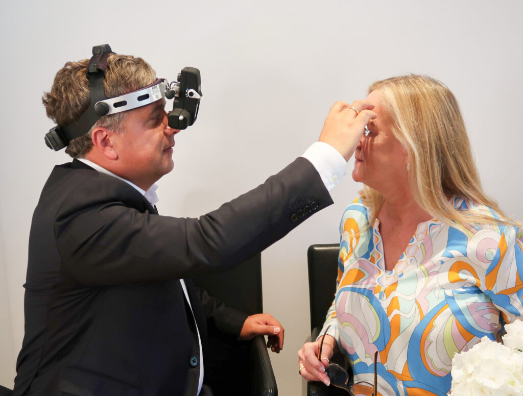
Funduscopy (ophthalmoscopy): The classic examination of the back of the eye by Dr. med. Mareen Schmidt for direct visual assessment of the retinal structure.
Three-mirror contact glass (if required): A specialized examination method that is used when necessary to also examine the outermost areas of the retina (periphery) precisely and in detail, for example in the case of suspected retinal tears.
Diseases of these structures often develop gradual and painless, but lead to irreversible damage if left untreated:
| Disease | Affected structure | Brief description | Symptoms (late stage) |
| Age-related macular degeneration (AMD) | Macula | Degeneration of the light-sensitive cells in the macula (dry and wet form). | Blurred vision, distorted vision (metamorphopsia), dark spot in the center. |
| Diabetic retinopathy | Peripheral and central retina | Damage to the retinal vessels due to high blood sugar. Leads to bleeding and swelling (macular edema). | Gradual deterioration of vision, Macular edema (loss of vision in the center). |
| Retinal detachment | Entire retina | Detachment of the retina from its supply layer. A Emergency! | Flashes of light, soot rain (black dots), black curtain in the field of vision. |
| Epiretinal gliosis (macular pucker) | Macular surface | Formation of a fine membrane on the macula that draws wrinkles. | Light Distortions (wavy lines), reduced visual acuity. |
The health of the retina is crucial for central vision. However, many serious diseases of this fine layer of tissue - from the age-related macular degeneration (AMD) up to the diabetic retinopathy - often develop gradually, without initial pain or noticeable symptoms. On the occasion of [current event, e.g. Day of Vision], [name of your practice/organization] points out the need for an early and comprehensive ophthalmological examination.
Affected structure: Macula
Brief description: Degeneration of the light-sensitive cells in the macula (dry and wet form).
Symptoms (late stage): Blurred vision, distorted vision (metamorphopsia), dark spot in the center.
Affected structure: Peripheral and central retina
Brief description: Damage to the retinal vessels due to high blood sugar. Leads to bleeding and swelling (macular edema).
Symptoms (late stage): Gradual deterioration of vision, Macular edema (Loss of vision in the center).
Affected structure: Entire retina
Brief description: Detachment of the retina from its supply layer. An emergency!
Symptoms (late stage): Flashes of light, Soot rain (black dots), Black curtain in the field of vision.
Affected structure: Macular surface
Brief description: Formation of a fine membrane on the macula that draws wrinkles.
Symptoms (late stage): Light Distortions (wavy lines), reduced visual acuity.
The Retina is the entire, large light-sensitive layer in the back of the eye. The Macula (yellow spot) is only a small, central area of the retina. However, the macula is the most important area, as it is responsible for the sharp vision, detailed vision and color vision is responsible.
Sudden symptoms such as Flashes of light (as if you were seeing a flash of lightning), a strong Soot rain (lots of black dots flying around) or an externally spreading black curtain in the visual field may indicate a retinal detachment. This is a ophthalmologic emergency and must be treated immediately.
The retina itself has no pain receptors. This is why most diseases such as Macular degeneration or the diabetic retinopathy painless. Pain in the eye is usually an indication of inflammation of the cornea, conjunctiva or increased intraocular pressure (glaucoma attack).
Yes, a healthy lifestyle is important. For the macula in particular, the intake of certain Antioxidants (e.g. lutein, zeaxanthin, vitamin C/E) and Omega-3 fatty acids slow down the progression of existing macular degeneration. Talk to us about special Food supplements to.
Yes, you can influence the development and progression of many Retinal diseases, in particular the Age-related macular degeneration (AMD) and the diabetic retinopathy, through your diet significantly influence.
Dry AMD: The more common, slowly progressing form. It is characterized by deposits (drusen) and gradual breakdown of photoreceptors.
Moist AMD: The rarer but more dangerous form. Here, new, leaky blood vessels grow under the retina, which release fluid and blood and can quickly lead to severe vision loss.


Check-ups, laser medicine, interdisciplinary ophthalmology

Retina, macular therapy, laser medicine, prevention & more
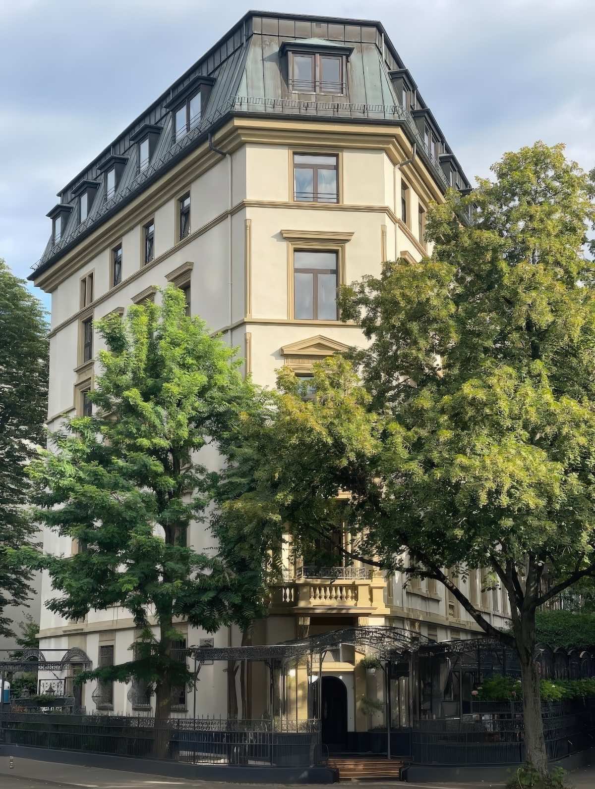
Address for the GPS:
Grüneburgweg 95 (corner of Liebigstraße), 60323 Frankfurt am Main.
Parking recommendations:
We recommend the following parking garages in the immediate vicinity:
Palmengarten parking garage: (Entrance via Siesmayerstraße). From there, it's about a 5-7 minute walk to the practice.
Campus Westend parking garage: (Entrance via Theodor-W.-Adorno-Anlage). Perfectly located for reaching the practice on foot in just a few minutes.
By bus (line 75):
Use the Bus route 75 and get off at the stop Eppsteiner Straße From there, it's only about a 2-3 minute walk (approx. 200 meters) along Grüneburgweg to the practice at the corner of Liebigstraße. Alternatively, you can use the bus stop. University Campus Westend to use.
By subway (U6 / U7):
Drive to the station Westend. Take the exit towards Feuerbachstraße/Liebigstraße. If you follow Liebigstraße straight ahead to the north, you will walk directly towards Villa Westend on Grüneburgweg (approx. 7-minute walk).
Alternative via the Palm Garden:
From the subway station Westend or the bus stop Palmengartenstrasse You can reach us after a short, pleasant walk past the Palm Garden.