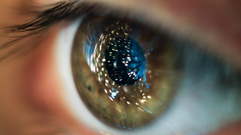
A retinal vascular occlusion, often referred to as an "eye stroke" or "retinal thrombosis," is a serious ophthalmological emergency that leads to a sudden, painless, and often dramatic deterioration of vision. It requires immediate and highly specialized evaluation, as every minute can be crucial for preserving your eyesight. At our ophthalmology practice in Frankfurt Westend, Dr. Mareen Schmidt, an experienced retinal specialist, focuses on determining the cause and extent of a retinal vascular occlusion. Retinal vascular occlusion to diagnose quickly and immediately initiate the correct, vision-preserving therapeutic measures.
Retinal vascular occlusion: What exactly happens in the eye?
To a Retinal vascular occlusion To understand this, one can imagine the blood supply to the retina as a road system. There are arteries that bring fresh, oxygen-rich blood to the retina (the "entry roads"), and veins that carry the deoxygenated blood away (the "exit roads"). A blockage can occur in either system, leading to completely different problems.
- Arterial occlusion: If an artery becomes blocked, the supply of oxygen and nutrients stops immediately. The affected retinal cells suffer irreversible damage within a very short time. This is the actual "eye stroke" and an absolute emergency.
- Venous occlusion: If a vein is blocked, blood pools in the retina because it can no longer drain away. This leads to bleeding, oxygen deprivation, and, most importantly, swelling of the central retina (macular edema), which threatens central vision.
Symptoms of a retinal vascular occlusion: An ophthalmological emergency
The symptoms of Retinal vascular occlusion They are almost always one-sided and painless, which makes them particularly insidious. Never ignore these warning signs!
If you experience any of the following symptoms, consult an ophthalmologist or eye clinic immediately:
- Sudden, severe loss of vision: Vision in one eye deteriorates dramatically within seconds or minutes, often like a curtain falling. This is typical of an arterial occlusion.
- A shadow or a "wall" in the field of vision: You notice a dark, motionless shadow moving into the image from one side. This is characteristic of a branch retinal vein occlusion.
- Generally blurred or foggy vision: The entire field of vision in one eye is blurred, as if through a dense fog. This often indicates a central retinal vein occlusion.
Any sudden, painless deterioration of vision is a warning sign that requires immediate investigation of a possible underlying condition. Retinal vascular occlusion requires.
Diagnosis of retinal vascular occlusion in our Frankfurt practice
If you suspect you have a Retinal vascular occlusion When you come to our practice, a fast and precise diagnosis is our top priority. We use state-of-the-art technologies to assess the extent of the damage and create the basis for an immediate treatment decision.
- Medical history and visual acuity test: We will ask about the exact time and type of visual impairment and measure your eyesight.
- Examination of the fundus: After dilating the pupil, Dr. Mareen Schmidt can directly assess the retina and identify the typical signs of a Retinal vascular occlusion such as bleeding, swelling, or a pale retina.
- Optical coherence tomography (OCT): This high-resolution scanning procedure is crucial for accurately measuring swelling of the macula (macular edema), the most common vision-threatening complication.
- Fluorescein angiography (FAG): This contrast-enhanced examination allows us to visualize the blood flow in the retinal vessels, pinpoint the exact location of the occlusion, and assess the extent of oxygen deficiency.
Retinal vascular occlusion: Arterial occlusion vs. venous occlusion in comparison
Although both fall under the term Retinal vascular occlusion The causes, urgency, and treatment options vary greatly depending on the specific case. The following table provides an overview.
|
Feature |
Arterial occlusion („eye infarction“) |
Venous occlusion („thrombosis“) |
|
Main problem |
Insufficient blood supply, acute oxygen deficiency. |
Impaired blood flow, blood congestion, swelling. |
|
Symptoms |
Mostly sudden, complete and dramatic loss of vision. |
Blurred vision, shadows; the extent depends on the location of the shutter. |
|
urgency |
Extremely high! Every minute counts; the window of opportunity for therapy is very small (a few hours). |
High. A timely presentation (within 1-2 days) is crucial. |
|
Therapy in the acute stage |
Attempt to dissolve the clot (e.g., through eye massage, pressure reduction, thrombolysis therapy in the clinic). |
Primarily no acute therapy, focus on treating complications. |
|
Main complication |
Irreversible destruction of the photoreceptor cells. |
Chronic macular edema, growth of abnormal blood vessels (neovascularization). |
|
Long-term therapy |
Prevention of re-occlusion. |
IVOM therapy (intraocular injection) against macular edema, laser therapy. |

Modern therapy for retinal vascular occlusion
The treatment of a Retinal vascular occlusion Treatment depends on the type of occlusion and any complications that arise. While options are limited in the case of an arterial occlusion, we can now treat the vision-threatening consequences of a venous occlusion very effectively.
- IVOM therapy (eye injection): The main complication after a retinal vein occlusion is swelling of the macula (macular edema). The treatment of choice is the injection of medication (VEGF inhibitors or cortisone) directly into the eye. This seals the blood vessels and reduces the swelling.
- Laser therapy (photocoagulation): If the oxygen deficiency in the retina is so severe that new, abnormal blood vessels begin to grow, these areas must be treated with a laser to prevent severe bleeding or a significant increase in pressure.
The crucial role of interdisciplinary clarification
A Retinal vascular occlusion is almost never just an eye problem. It is a strong warning sign of an underlying systemic cardiovascular disease. Therefore, after acute ophthalmologic care, an immediate and thorough internal or cardiologic workup is essential to find the cause and prevent future, potentially life-threatening events such as a stroke or heart attack. As your ophthalmologist, we will initiate these important steps and work closely with your family doctor.
Frequently asked questions about retinal vascular occlusion
Here you will find answers to common questions that patients ask us about Retinal vascular occlusion in our Frankfurt practice.
Is a retinal vascular occlusion the same as a stroke?
A Retinal vascular occlusion It is essentially a "stroke in the eye." The mechanisms and risk factors (high blood pressure, diabetes, high cholesterol, cardiac arrhythmias) are the same. Therefore, an ocular infarction must be taken just as seriously as a stroke in the brain and requires a comprehensive internal medical evaluation for prevention.
Can I become completely blind due to a retinal vascular occlusion?
Yes, unfortunately, that's possible. A complete central retinal artery occlusion leads in most cases to very severe, permanent visual impairment, even to the point of practical blindness in the affected eye. A severe central retinal vein occlusion can also lead to massive vision loss if left untreated. Rapid diagnosis and treatment after a Retinal vascular occlusion is therefore crucial.
Is the treatment (eye injection) painful?
No. IVOM therapy after a Retinal vascular occlusion The procedure is performed using our virtually painless 5-S method. Thanks to intensive local anesthesia with eye drops, the injection itself is not painful. You will only feel a brief, slight pressure, which makes the treatment very well tolerated.
How often are injections necessary after a retinal vessel occlusion?
Treatment of macular edema after a Retinal vascular occlusion It usually requires a series of injections. Initially, monthly treatments are often necessary. Once the swelling has subsided, the treatment intervals are individually extended to maintain the condition with as few injections as possible.
Can I prevent a retinal blood vessel occlusion?
Yes, the best prevention is the consistent control and treatment of the underlying risk factors. Good control of your blood pressure, blood sugar, and blood lipid levels is the most effective protection. If you are a smoker, quitting smoking is the single most important measure to reduce your risk of developing [a specific type of cancer]. Retinal vascular occlusion to lower.
Can my vision recover after a retinal vascular occlusion?
The prognosis is highly individual. After an arterial occlusion, the chance of recovery is unfortunately low. After a venous occlusion, the prognosis depends heavily on the extent of the occlusion and the success of treating the macular edema. With modern intravitreal injection (IVOM) therapies, a Retinal vascular occlusion A significant improvement in eyesight can often be achieved if treatment begins early.
I have high blood pressure. Am I at particular risk?
Yes, high blood pressure is the most important risk factor for a Retinal vascular occlusion. It leads to hardening and damage of the artery walls, which can cause compression and thus blockage at the points where they cross with the veins. Regular eye examinations are therefore an important preventative measure for every patient with high blood pressure.
Why do I need to see my family doctor or cardiologist after the diagnosis?
A Retinal vascular occlusion This is a wake-up call. It indicates that your blood vessels throughout your body are at risk. A comprehensive internal medical examination is necessary to determine the cause (e.g., undetected high blood pressure, cardiac arrhythmia, narrowing of the carotid artery) and to initiate targeted therapy to prevent heart attack and stroke.
How long will it take for my vision to return to normal after the treatment?
The healing process after a Retinal vascular occlusion The process is lengthy. Reducing swelling with IVOM therapy can take several weeks to months. A gradual improvement in vision is often observed over an extended period. Patience and consistent adherence to treatment appointments are crucial for success.
Why should I go to a retina specialist like Dr. Mareen Schmidt?
The diagnosis and management of a Retinal vascular occlusion This requires the highest level of expertise and access to state-of-the-art diagnostic and therapeutic procedures. As an experienced retinal specialist, Dr. Mareen Schmidt can quickly and reliably assess the situation, perform all necessary therapies (IVOM, laser) at the highest level, and coordinate the crucial interdisciplinary collaboration.
Act quickly if you experience sudden vision problems.
A Retinal vascular occlusion It is a serious threat to your eyesight, but modern ophthalmology offers effective treatment options, especially in the case of a retinal vein occlusion. The most important thing is not to waste any time. If you notice a sudden, painless deterioration of vision in one eye, don't hesitate. Schedule an appointment immediately for a comprehensive examination and consultation at our Frankfurt practice.