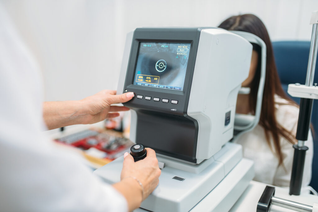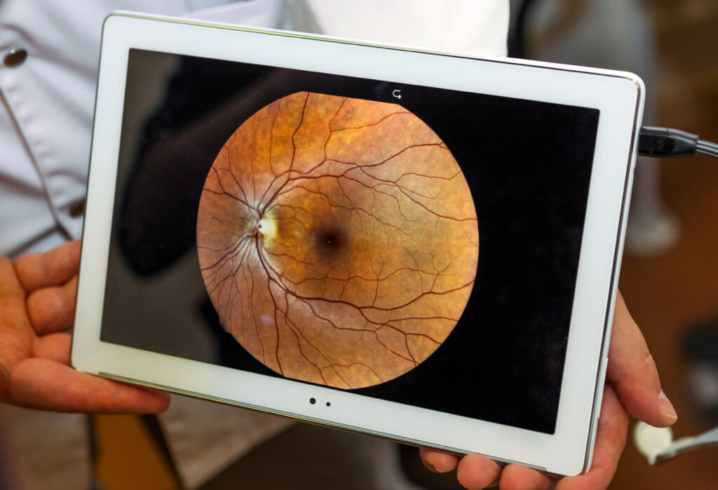
Wide-angle ophthalmometry is one of the most advanced and patient-friendly methods in modern ophthalmology, enabling a comprehensive and precise assessment of your retinal health. This revolutionary technology allows us to capture a wide area of your fundus in a single, high-resolution image – and in the vast majority of cases, without the need to dilate your pupils with medication. At the ophthalmology practice of Dr. Mareen Schmidt in Frankfurt's Westend district, this technique is available. Wide-angle photography of the eye a central component of our careful preventative care philosophy, which focuses on maximum diagnostic certainty with the highest level of patient comfort.
What is wide-angle photography of the eye and how does it work?
The Wide-angle photography of the eye, Fundus photography, often referred to as ultra-wide-angle retinal imaging, is a non-contact scanning procedure that creates a digital panoramic image of your retina in seconds. You simply look into the device for a brief moment and focus on a light. A harmless laser then scans the back of the eye, generating an image that covers up to 200 degrees, or approximately 801 TP3T, of your entire retina. This is a far larger area than could ever be seen during a conventional ophthalmoscope examination without pupil dilation. The result is a crystal-clear digital "photo" that Dr. Mareen Schmidt can immediately analyze on screen, enlarge, and save for future comparisons.
Wide-angle photography of the eye and its decisive advantages
The Wide-angle photography of the eye It has revolutionized retinal screening for both patients and doctors. The advantages over the classic examination method with medication-induced pupil dilation are immense and make this important preventative care simpler than ever before.
Your advantages at a glance:
- No glare sensitivity: Since pupil-dilating drops are unnecessary in most cases, you will not be blinded by bright light for hours after the examination.
- No driving ban: You will be fully fit to drive immediately after the examination and can continue your daily life without restrictions.
- Speed and comfort: The procedure itself takes only a few seconds and is completely contactless and painless. There is no long waiting time for the drops to take effect.
- Highest precision and documentation: The high-resolution digital image enables precise analysis of even the smallest changes in the retina. Storing the images allows for objective comparison over years, which is essential for monitoring disease progression.
- Comprehensive overview: The Wide-angle photography of the eye It also captures the outer (peripheral) areas of the retina, which are difficult to see during a standard examination.
Wide-angle photography of the eye in Frankfurt: For which diseases is it crucial?
The Wide-angle photography of the eye is a universal preventative tool that plays a crucial role in the early detection of a variety of serious eye diseases.
The examination is particularly important for assessing:
- Diabetic retinopathy: For the detection and documentation of vascular changes, bleeding, or oxygen-deficient areas caused by diabetes.
- Retinal changes in high myopia (nearsightedness): In nearsighted eyes, the retina is often stretched and more prone to developing thin spots, holes, or tears in the periphery. Wide-angle photography of the eye can represent these areas excellently.
- Age-related macular degeneration (AMD): For the documentation of deposits (drusen), hemorrhages, membranes, pigment changes and for the assessment of the condition of the macula as a whole.
- Retinal changes in high blood pressure: To assess the condition of the fine blood vessels in the back of the eye, which allows conclusions to be drawn about the entire vascular system.
- Retinal tumors or moles (nevus): For the precise assessment of the size and progression of pigment changes in the back of the eye.
- Vascular occlusions: To document the extent of a "thrombosis" in the eye (bleeding, avascular areas, complications of reduced blood flow)
Wide-angle photography of the eye compared to other methods
Modern ophthalmology offers various procedures for examining the retina. Wide-angle photography of the eye It has established itself as a central instrument for preventative measures.
|
Examination procedures |
Main application / focus |
The need for pupil dilation |
|
Classical ophthalmoscopy |
Basic examination of the central fundus. |
Often necessary for a good overview. |
|
Wide-angle photography of the eye |
Comprehensive screening of the entire retina, ideal preventative care and documentation. |
Usually not necessary. |
|
Optical Coherence Tomography (OCT) |
Micrometer-precise layered image of the central retina (macula). |
Usually not necessary, but often advantageous. |
Frequently asked questions about wide-angle photography of the eye
Here you will find answers to frequently asked questions about this modern preventive examination in our Frankfurt practice.
Is wide-angle photography of the eye just as good as an examination with dilating drops?
For most preventive examinations and screening purposes, the Wide-angle photography of the eye This provides an excellent and often even more comprehensive overview of the retina than a conventional examination. Digital documentation also allows for superior monitoring of disease progression. Only in very specific cases, with a very small pupil, or when a therapeutic intervention is planned, might dilating drops be necessary in addition.
Does the test work for every patient?
In the vast majority of cases, it works Wide-angle photography of the eye No problems. In patients with an extremely small pupil or very advanced cataracts, image quality may be limited. In these rare cases, it may be necessary to dilate the pupil further to ensure a reliable assessment and complete the examination.
Is wide-angle photography of the eye painful?
No, the Wide-angle photography of the eye It is completely painless. You will feel nothing except a brief, harmless flash of light, similar to a camera. The entire process is contactless and only takes a few seconds, making it a very comfortable and stress-free examination method for patients of all ages.
How long does the examination take?
The actual recording for the Wide-angle photography of the eye The procedure takes only a few seconds per eye. The entire appointment, including preparation and the subsequent discussion of the images with Dr. Mareen Schmidt, is usually completed within 15-20 minutes. This makes this important retinal screening extremely time-efficient and easy to integrate into everyday life.
Will my health insurance cover the costs of wide-angle photography of the eye?
The Wide-angle photography of the eye This is a modern, private medical service, the costs of which are covered by private health insurance. The advantages in terms of comfort and diagnostic accuracy convince most patients.
Why is wide-angle photography of the eye so important for nearsightedness?
In nearsighted people, the eyeball is longer, which leads to a stretching of the retina. This increases the risk of developing thin spots, holes, or tears in the peripheral retina. Wide-angle photography of the eye It enables an excellent representation of these often difficult-to-see peripheral areas and is therefore an ideal method for preventative care in cases of high myopia.
Can the examination also detect glaucoma?
Yes, indirectly. The high-resolution recording at the Wide-angle photography of the eye It also provides a very detailed image of the optic nerve head. An experienced ophthalmologist like Dr. Mareen Schmidt can already detect early signs of glaucomatous damage here. However, for a complete glaucoma diagnosis, additional measurements such as intraocular pressure and an OCT scan of the optic nerve are necessary.
How often should I have a wide-angle photograph of my eye taken?
We recommend regular Wide-angle photography of the eye As part of your general eye check, it is strongly recommended if you have risk factors such as diabetes, high blood pressure or high myopia, in order to safely monitor the retina.
Is there a risk of radiation exposure during the examination?
No, with the Wide-angle photography of the eye A harmless, visible laser light with very low energy is used. There is absolutely no X-ray or UV radiation. The examination is completely safe and can be repeated as often as necessary, even for children or during pregnancy, without any concerns.
Why should I have the examination done at your practice in Frankfurt?
At Dr. Mareen Schmidt's practice, you benefit from the combination of state-of-the-art technology and the highest level of professional expertise: the necessary correct interpretation of the images. We take the time to show you the high-resolution images of your retina and to explain the findings, along with their sound interpretation, in a way that is easy to understand. Wide-angle photography of the eye With us, it's more than just a photo – it's the basis of comprehensive, personal health advice.
See your retirement planning in a new light.
The Wide-angle photography of the eye This technology revolutionizes eye exams, making them easier, faster, and more comfortable than ever before. There's no longer any reason to postpone that important fundus examination. Take advantage of this modern technology at our Frankfurt practice. Schedule an appointment with Dr. Mareen Schmidt for your comprehensive and thorough eye exam today.